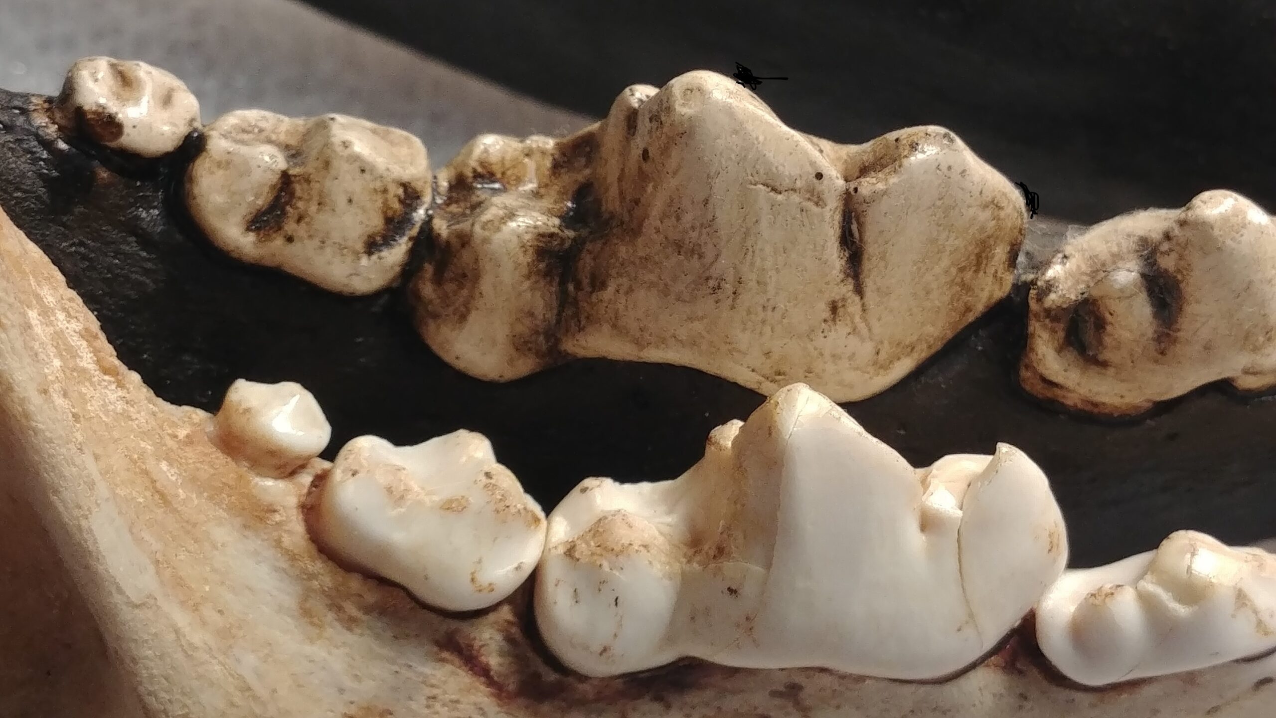
The head of the femur is just one of many skeletal epiphyses in the body. An epiphysis is a separate ossification center which is present at one or both ends of some bones, and is where growth in length of a bone takes place. If you look at the bottom of this femoral head (photo), you can see that the surface has a roughened texture. This is where the epiphyseal disk is found, a layer of cartilage between the epipiysis and bone shaft, and this is where growth occurs. Once full adult skeletal growth is completed, the epiphyses will fuse to the main shaft of the bone and you’ll have one complete bone. Since this mastodon femoral head is unfused, we can infer that the animal was a sub-adult. It’s still one big mastodon; it’s adult size but not yet skeletally mature.
Speaking of femoral heads, at the last museum that I was with we had a large colony of flesh-eating beetles, called dermestid beetles, that we used to clean flesh off of skeletal specimens (more about that in upcoming blogs!). One of the graduate students that I worked with had unfortunately developed a severe case of arthritis, very unusual for someone as young as her. As part of the disease, the protective cartilage on one of her femoral heads had worn away and was causing her great pain, thus the head of her femur had to be surgically removed. After the surgery she hobbled on her crutches into the museum one day and said to me, “Dave, if I can get the femoral head from the surgery, would you put it into the dermestid colony and clean it up for me!?” I had used the colony several times to clean human bones from forensic cases brought in by the State Crime Lab, so I knew the beetles would clean human tissue. So I said “Sure, if you can get the specimen from your doctors go ahead and bring it in.” She then proceeded to reach into her backpack and pull out a small jar containing, you guessed it, her femoral head! I was surprised at first, but having worked with forensic science grad students for so long I really wasn’t too shocked!